Home
/ Foot And Leg Bones Diagram, Bones The Of Foot Stock Vector Illustration Of Phalanges 131402653, Sits over the front of the knee joint.
Foot And Leg Bones Diagram, Bones The Of Foot Stock Vector Illustration Of Phalanges 131402653, Sits over the front of the knee joint.
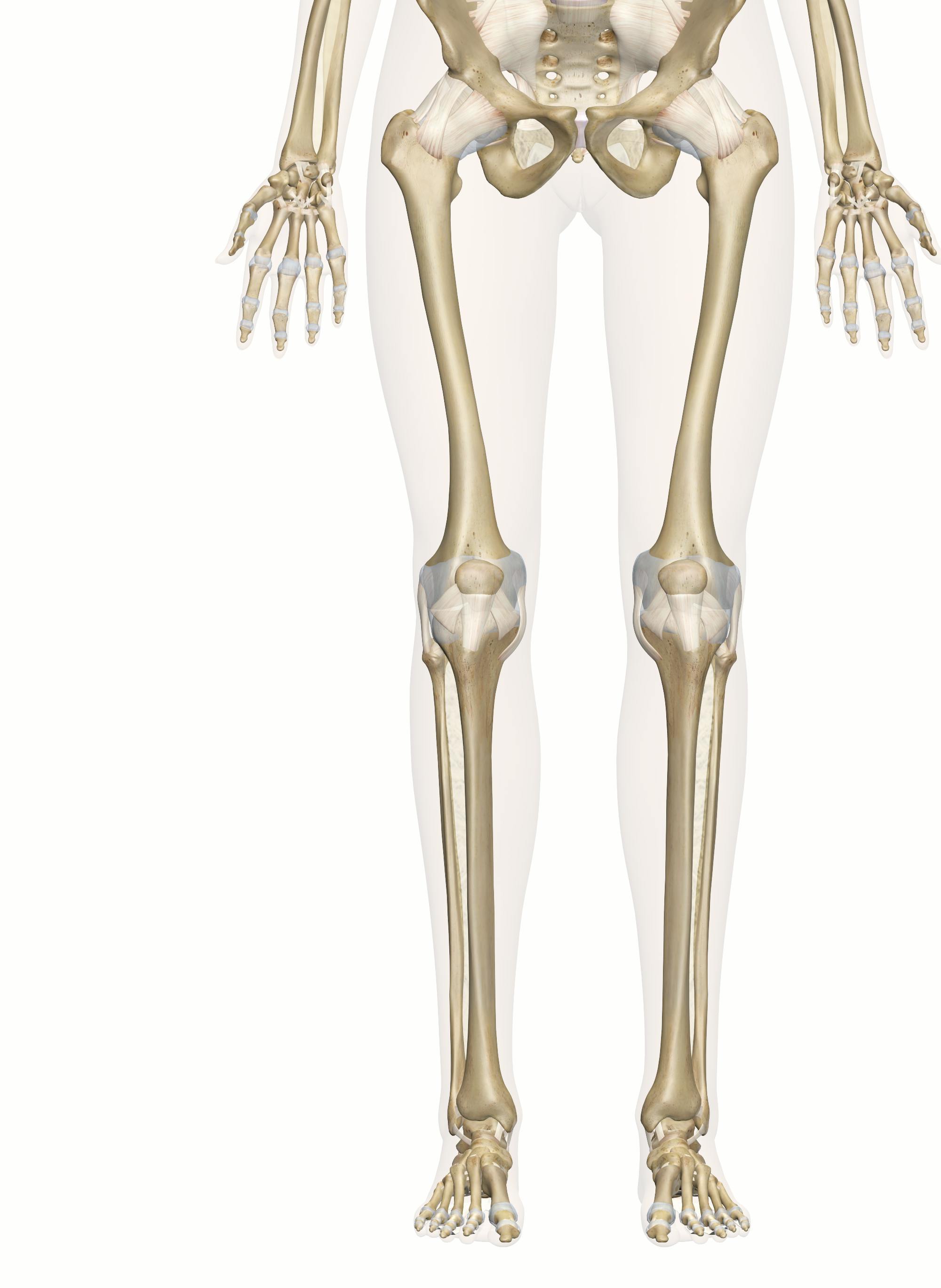
Foot And Leg Bones Diagram, Bones The Of Foot Stock Vector Illustration Of Phalanges 131402653, Sits over the front of the knee joint.. The bones of the leg and foot form part of the appendicular skeleton that supports the many muscles of the lower limbs. The seven tarsal bones are: The calcaneus (heel bone) is the largest bone in the foot. The leg is specifically the region between the knee joint and the ankle joint. Lower limb blood vessel coloring page, lower limb muscles anatomy worksheet and foot and ankle bones unlabeled are three of main things we want to present to you based on the gallery title.
Like already mentioned, the hindfoot is the posterior part of the foot. The lower limb contains 30 bones. The smaller lateral bone of the lower leg. Lastly, this diagram shows the lateral aspect of the foot, with lateral meaning to the side. this is the view of the foot from the side of the body, then; Fpe medical review board a foot pain diagram is a great tool to help you work out what is causing your ankle and foot pain.
Bones Of The Leg And Foot Interactive Anatomy Guide from innerbody.imgix.net It connects with the tibia and fibula bones of the lower leg. Chloe wilson bsc(hons) physiotherapy reviewed by: The bones of the leg and foot form part of the appendicular skeleton that supports the many muscles of the lower limbs. Use lucidchart to visualize ideas, make charts, diagrams & more. Sits over the front of the knee joint. Ankle & lower leg anatomy. The knee is a strong but flexible hinge joint. There are a whole range of structures e.g.
Your leg bones are the longest and strongest bones in your body.
The calcaneus, or heel bone: The knee is a strong but flexible hinge joint. The thigh bone or longest bone in the body. Like already mentioned, the hindfoot is the posterior part of the foot. Distal to the ankle is the foot. The talus bone supports the leg bones (tibia and fibula), forming the ankle. It extends from your knee joint upwards to the ankle joint downwards. The bones of your leg and foot helped give you the ability to score that field goal. The lower limb contains 30 bones. Is the inner and larger of the 2 lower leg bones, extending from the knee to the ankle. These bones articulate (connect) to the talus or ankle bone at the tibiotalar joint (ankle joint) allowing the foot to move up and down. The medial, larger bone of the lower leg. As these nerves descend toward the thighs, they form two networks of crossed nerves known as the lumbar plexus and sacral plexus.
It extends from your knee joint upwards to the ankle joint downwards. The talus bone supports the leg bones (tibia and fibula), forming the ankle. The bones of the foot provide mechanical support for the soft tissues; Is the outer and narrower of the 2 lower leg bones, extending from the knee to. The medial, larger bone of the lower leg.
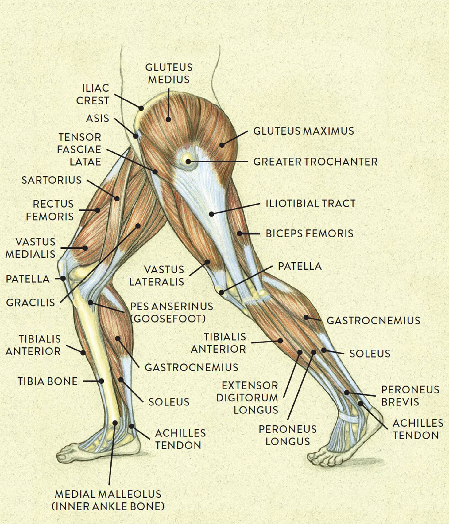
Muscles Of The Leg And Foot Classic Human Anatomy In Motion The Artist S Guide To The Dynamics Of Figure Drawing from doctorlib.info A foot bone that sits above the heel bone (talus). Your leg bones are the longest and strongest bones in your body. The anatomy of the leg and foot bones. Tibia and fibula (long bones) the foot is connected to the body where the talus articulates with the tibia and fibula. Distal to the ankle is the foot. At the same time, the bones and joints of the leg and foot must be strong enough to support the body's weight while remaining. Helping the foot withstand the weight of the body whilst standing and in motion. As these nerves descend toward the thighs, they form two networks of crossed nerves known as the lumbar plexus and sacral plexus.
The knee is a strong but flexible hinge joint.
Bones of the leg and foot, lower leg bone anatomy, leg bones anatomy, leg muscles, leg bones diagram, leg bone structure, leg anatomy muscles, parts of the lower leg. It connects with the tibia and fibula bones of the lower leg. Tibia and fibula (long bones) the foot is connected to the body where the talus articulates with the tibia and fibula. The view of the part of the foot that faces outward. Moveable tissue) surround the true ankle and subtalar joints, binding the bones of the leg to each other and to those of the foot. At the same time, the bones and joints of the leg and foot must be strong enough to support the body's weight while remaining. Helping the foot withstand the weight of the body whilst standing and in motion. They can be divided into three groups: While we talk concerning leg anatomy worksheets, we've collected particular similar images to give you more ideas. The talus bone supports the leg bones (tibia and fibula), forming the ankle. Lastly, this diagram shows the lateral aspect of the foot, with lateral meaning to the side. this is the view of the foot from the side of the body, then; Leg, ankle and foot bones. It forms the bottom of the ankle joint, articulating with the tibia and fibula (shin bones) and the top of the subtalar joint, articulating with the calcaneus (heel bone).
Fpe medical review board a foot pain diagram is a great tool to help you work out what is causing your ankle and foot pain. Distal to the ankle is the foot. The nerves of the leg and foot arise from spinal nerves connected to the spinal cord in the lower back and pelvis. These are the femur, patella, tibia, fibula, tarsal bones, metatarsal bones, and phalanges (see figure 6.51). Like already mentioned, the hindfoot is the posterior part of the foot.
Anatomy Of The Foot And Ankle Orthopaedia from orthopaedia.com Though often thought of as a problem of the feet or legs, lameness can involve virtually any part of the body and can originate in bone or soft tissue. Lastly, this diagram shows the lateral aspect of the foot, with lateral meaning to the side. this is the view of the foot from the side of the body, then; Its main function is to allow for plantar flexion and dorsiflexion of the foot. The ankle joint is both a synovial joint and a hinge joint. While we talk concerning leg anatomy worksheets, we've collected particular similar images to give you more ideas. The nerves of the leg and foot arise from spinal nerves connected to the spinal cord in the lower back and pelvis. Tibia and fibula (long bones) though the tibia (commonly called the shin bone) is not a part of the foot, it plays an important role. This bone creates the lower portion of the ankle joint.;
Tibia and fibula (long bones) the foot is connected to the body where the talus articulates with the tibia and fibula.
It connects with the tibia and fibula bones of the lower leg. Tibia and fibula (long bones) though the tibia (commonly called the shin bone) is not a part of the foot, it plays an important role. The ankle consists of three bones attached by muscles, tendons, and ligaments that connect the foot to the leg. The smaller lateral bone of the lower leg. The view of the part of the foot that faces outward. A foot bone that sits above the heel bone (talus). Ankle & lower leg anatomy. The ankle is a joint that connects the lower leg to the foot. The knee joint is the largest joint in the body and is primarily a hinge joint, although some sliding and rotation occur. The medial, larger bone of the lower leg. Let's review all of these bones one last time. Learn vocabulary, terms and more with flashcards, games and other study tools. Your leg bones are the longest and strongest bones in your body.
At the same time, the bones and joints of the leg and foot must be strong enough to support the body's weight while remaining leg bones diagram. The ankle is made off the tibia and.
